

What is the difference between a high - magnification and a low - magnification eyepiece for a compound microscope?
As a dedicated supplier of microscope eyepieces, I've witnessed firsthand the diverse needs of researchers, educators, and hobbyists when it comes to compound microscopes. One of the most fundamental decisions users face is choosing between high - magnification and low - magnification eyepieces. In this blog, I'll delve into the differences between these two types of eyepieces, exploring their characteristics, applications, and how they can impact your microscopy experience.
Magnification and Field of View
The most obvious difference between high - magnification and low - magnification eyepieces lies in their magnification power. Low - magnification eyepieces, such as the WF10X 18mm Eyepiece, typically offer magnifications in the range of 5X to 10X. These eyepieces provide a wider field of view, allowing users to observe a larger area of the specimen at once. This is particularly useful when initially locating a specimen on the slide or when examining large - scale features.

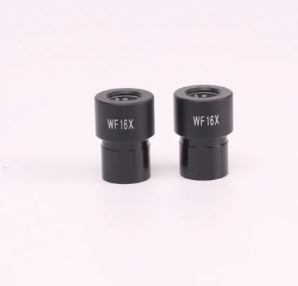
On the other hand, high - magnification eyepieces, like the WF16X Eyepiece, offer magnifications of 15X or higher. They provide a more detailed view of the specimen but at the cost of a narrower field of view. With high - magnification eyepieces, users can focus on specific features or structures within the specimen, making them ideal for detailed analysis and study.
Depth of Field
Another important difference is the depth of field, which refers to the range of distance within the specimen that appears in sharp focus. Low - magnification eyepieces generally have a greater depth of field. This means that more of the specimen will be in focus at the same time, even if the specimen is not perfectly flat. This is beneficial when observing specimens with uneven surfaces or when trying to get an overall view of a thick specimen.
In contrast, high - magnification eyepieces have a shallower depth of field. Only a very thin slice of the specimen will be in focus at any given time. This requires more precise focusing and can make it challenging to observe specimens with significant depth. However, the shallow depth of field can also be an advantage when trying to isolate and examine specific layers or structures within a specimen.
Image Brightness
Image brightness is also affected by the magnification of the eyepiece. Low - magnification eyepieces typically allow more light to reach the eye, resulting in a brighter image. This is because they have a larger exit pupil, which is the diameter of the beam of light that emerges from the eyepiece. A larger exit pupil means that more light can enter the eye, making the image appear brighter.
High - magnification eyepieces, on the other hand, have a smaller exit pupil. As a result, less light reaches the eye, and the image may appear dimmer. To compensate for this, users may need to increase the illumination of the microscope or use a more powerful light source.
Eye Relief
Eye relief is the distance between the eyepiece lens and the user's eye at which the entire field of view can be seen. Low - magnification eyepieces often have greater eye relief. This is especially important for users who wear glasses, as it allows them to comfortably view the specimen without having to remove their glasses.
High - magnification eyepieces usually have shorter eye relief. This can be a drawback for glasses wearers, as they may have to remove their glasses to get a clear view of the entire field of view. However, some high - magnification eyepieces are designed with adjustable eye cups or other features to accommodate glasses wearers.
Applications
The choice between high - magnification and low - magnification eyepieces depends largely on the specific application. Low - magnification eyepieces are commonly used in educational settings, where students are learning to use microscopes and need to get an overall view of the specimen. They are also useful for quick inspections and general observations, such as checking the quality of a sample or looking for large - scale features.
High - magnification eyepieces are essential in research and scientific applications, where detailed analysis of specimens is required. They are used to study cell structures, microorganisms, and other small - scale features. For example, in microbiology, high - magnification eyepieces are used to identify bacteria and other microorganisms based on their morphological characteristics.
Our Product Range
As a microscope eyepiece supplier, we offer a wide range of high - and low - magnification eyepieces to meet the diverse needs of our customers. Our H10X Eyepiece is a popular low - magnification option, providing a clear and bright image with a wide field of view. It is suitable for both educational and general laboratory use.
Our high - magnification WF16X Eyepiece is designed for detailed analysis and research. It offers excellent optical performance and a sharp, clear image, even at high magnifications.
We also have the WF10X 18mm Eyepiece, which combines a moderate magnification with a large field of view and good eye relief. This makes it a versatile option for a variety of applications.
Contact Us for Procurement
If you're in the market for microscope eyepieces, whether it's a high - magnification eyepiece for in - depth research or a low - magnification eyepiece for general observations, we're here to help. Our team of experts can provide you with detailed information about our products and assist you in choosing the right eyepiece for your specific needs. We offer high - quality products at competitive prices, and we're committed to providing excellent customer service.
Don't hesitate to reach out to us to start a procurement discussion. We look forward to working with you to enhance your microscopy experience.
References
- Microscopy: A Practical Approach, Third Edition, edited by David B. Murphy and Richard F. Murphy.
- Fundamentals of Light Microscopy and Electronic Imaging, by Douglas B. Murphy.
- Handbook of Biological Confocal Microscopy, Third Edition, edited by James B. Pawley.