

Microscope Eyepiece
Manufacturing Strength You Can Trust
Founded in 1965, Phenix Optical Stock Co., Ltd. has a history of over 50 years. It is one of the largest optical instrument manufacturers in China and the first publicly listed company in the optical industry. In July 2014, it was acquired by the China Electronics Technology Group (CETC). The company primarily manufactures microscopes, surveying instruments, cameras, telescopes, optical and video equipment, and optical lenses. Additionally, it offers comprehensive R&D and manufacturing capabilities, including precision metal machining, precision injection molding, and surface decoration.
Jiangxi Phenix Optical Technology Co., Ltd. is a subsidiary of Phenix Optical Stock Co., Ltd. As a renowned state-owned enterprise, it integrates the R&D and production of comprehensive optical instruments. Its main products include biological, fluorescence, phase contrast, stereo, metallurgical, polarizing, and digital microscopes, as well as interactive digital microscope laboratories. These professional microscopes are widely used in education, healthcare, industrial manufacturing, forestry, agriculture, animal husbandry, and environmental monitoring.


Microscope Eyepiece Production Process
Optical Material Selection & Pre-processing
The process begins with selecting high-quality optical glass (such as K9 or ZF2) to ensure high uniformity and proper refractive indices. The raw glass blanks undergo strict hardness checks. Before rough grinding, a "safety angle" is often machined on small lenses to prevent chipping during subsequent processing.

Precision Grinding & Centering
This stage focuses on achieving the required surface shape and strict center deviation (typically less than 0.1mm). Lenses are cemented to a fixture with a thin, uniform adhesive layer. The outer diameter is turned, and end runout is strictly controlled (within 0.05mm) using dial indicators to ensure perfect optical alignment.

Polishing & Surface Accuracy
Advanced polishing techniques (such as the 333 polishing method using three different hardness pads) are employed to achieve high surface accuracy. The process requires strict control over the number of fringes (e.g., N=1-2, ΔN=0.2-0.3) and local deviations, especially in the central zone, to guarantee clear imaging.

Cleaning & Cementing Assembly
For multi-element eyepieces (like Kellner or Plössl), individual lenses are cleaned in a dust-free environment to prevent air bubbles. UV-curable adhesives are applied, and the lenses are cured at a constant temperature to avoid uneven stress. The assembly is then left for 24 hours to release stress and stabilize.

Optical Coating
To maximize light transmission and reduce internal stray light or ghosting, lenses undergo coating processes (such as single-layer MgF2 or multi-layer broadband anti-reflection coatings). Before coating, lenses are cleaned using ultrasonic waves and IPA. The coating thickness is precisely controlled by observing color changes or using film thickness monitors.

Mechanical Housing Manufacturing
The metal housings and spacer rings are manufactured using precision CNC machining or wire-cut EDM. The flatness of spacer rings is strictly controlled (less than 0.1mm), and deep, dense anti-reflection threads are machined on the inner walls to minimize light scattering.

Final Assembly & Quality Testing
Lenses are assembled into the housing with uniform pressure (tightened diagonally). The final product undergoes rigorous testing, including star-point testing for astigmatism, high/low-temperature cycling (-40°C to 60°C), and matching tests for binocular microscopes (ensuring magnification and field of view differences are ≤1%).

Company Profile
Jiangxi Phenix Optical Technology Co., Ltd. Is the first listed company in the Chinese optical industry (SSE code: 600071), which is Successfully listed on the Shanghai Stock Exchange in 1997. It covers an area of about 333,000㎡ and employees about 3300 people.
We offer exclusive services you can't find with other companies. We have developed a unique service system designed to assist you in building your own Microscopes And of course our team members are always standing by to help you, Chat, Phone, or Email.
Why Choose Us
Professional Team
We offer exclusive services you can't find with other companies. We have developed a unique service system designed to assist you in building your own Microscopes And of course our team members are always standing by to help you, Chat, Phone, or Email.
Factory
Jiangxi Phenix Optical Technology Co., Ltd. Is the first listed company in the Chinese optical industry (SSE code: 600071), which is Successfully listed on the Shanghai Stock Exchange in 1997. It covers an area of about 333,000㎡ and employees about 3300 people.
Our Certificate
We always feel that all success of our company is directly related to the quality of the products we offer. They meet the highest quality requirements as stipulated in ISO9001, ISO14001, ISO45001 and SGS authentication and our stringent quality control system.
Production Equipment
We have a huge production workshop and production equipment, under the premise of ensuring quality can quickly complete the order production.
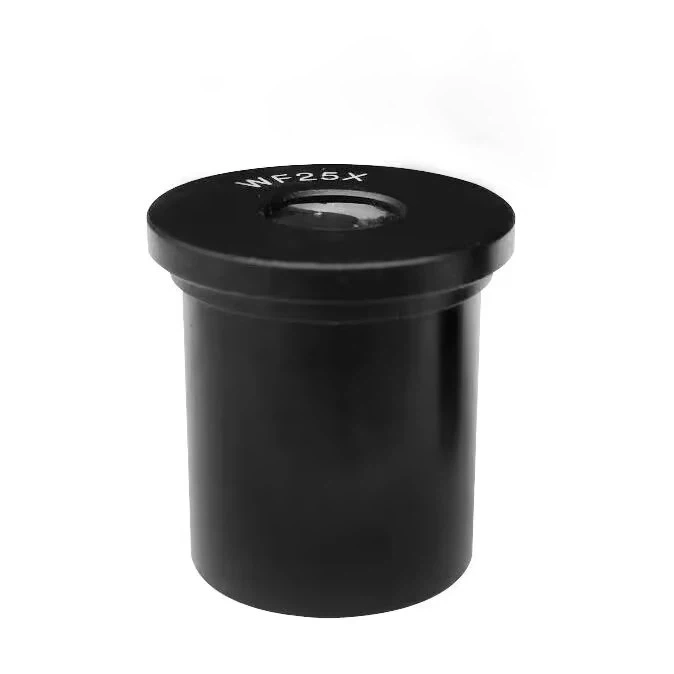
Microscope eyepieces are the lenses that you look through, near where you place your eye. The optical tube is what part of the microscope that holds the eyepiece. A compound microscope has one or more eyepieces that can be changed or swapped to other eyepieces, if they are compatible with the same tube diameter.
Its main function is to magnify the real image magnified by the objective lens again, thereby forming a clear virtual image at a clear viewing distance; therefore, its quality will ultimately affect the quality of the object image. In photomicrography, a real image is formed on the ground glass. In addition to magnifying, some eyepieces (such as compensating eyepieces) can also correct residual aberrations generated during the imaging process of the objective lens. The construction of eyepieces is much simpler than objective lenses. Because the light beam passing through the eyepiece is nearly parallel, spherical aberration and longitudinal (axial) chromatic aberration are not serious. Only horizontal chromatic aberration (amplified chromatic aberration) is considered when designing.
Benefits of Microscope Eyepiece
Easy to operate
The optical microscope has a relatively simple structure and is easy to use, suitable for teaching, primary research and routine testing.
Safe and non-destructive
The requirements for samples are relatively loose and will not be damaged by high-energy electron beams. It is suitable for observing living cells and tissue samples (especially under special techniques such as phase contrast and fluorescence).
Higher resolution
Although limited by the wavelength of light, it can still provide sufficient resolution to observe many details in the microscopic world, such as microorganisms, cell structures and certain fine particles.
Rich lighting and staining methods
A variety of light sources and staining techniques can be used to make sample features more distinct, which is helpful for morphological and functional research.
Type of Microscope Eyepiece
Micrometer eyepiece
A graduated glass piece is added to the eyepiece for quantitative measurement, or for measuring the length of microindentation. The scale can be designed on straight lines, cross lines, square grids, concentric circles or other geometric figures according to the purpose of measurement.
Photographic eyepiece
This eyepiece is specially used for photography or close-range projection, and cannot be used for microscopic observation or magnification alone. Its aberration correction is basically the same as that of the compensation eyepiece, and it should be used with a plan apochromatic objective lens or a semi-apochromatic objective lens to achieve a sufficiently flat image at the specified magnification.
Wide-angle eyepiece
Generally, the field of view angle of the eyepiece is about 30°. Wide-angle eyepieces refer to flat-field eyepieces with a field of view of more than 50° and a magnification of more than 12.5 times, and flat-field eyepieces with a field of view of more than 40° and a magnification of less than 10 times.
The eyepiece is a visual optical device used to observe the image formed by the front optical system. It is a component of visual optical instruments such as telescopes and microscopes. Its main function is to magnify the real image magnified by the objective lens again. In order to eliminate aberrations, the eyepiece is usually composed of several lenses, with a larger field of view and angle of view magnification.
The eyepiece is also the main component of the microscope. Its main function is to magnify the real image magnified by the objective lens again, thereby forming a clear virtual image at a clear viewing distance; therefore, its quality will ultimately affect the quality of the object image. In photomicrography, a real image is formed on the ground glass. In addition to magnifying, some eyepieces (such as compensating eyepieces) can also correct residual aberrations generated during the imaging process of the objective lens. The construction of eyepieces is much simpler than objective lenses.

An objective lens is a lens group composed of several lenses. The purpose of combined use is to overcome the imaging defects of a single lens and improve the optical quality of the objective lens. The magnifying effect of a microscope mainly depends on the objective lens. The quality of the objective lens directly affects the quality of the microscope image. It is the main component that determines the resolution and image clarity of the microscope, so the correction of the objective lens is very important.
The structure of the objective lens is complex and precision-made. It is usually composed of a lens group. Each lens is separated by a certain distance from each other to reduce the phase difference. Each set of lenses is made of one or several lenses of different materials and parameters glued together.
How to Choose the Right Eyepiece
Measure counting line
Eyepieces can be used to make measurements by adding a small disc-shaped glass reticle (sometimes called a counting line or reticle) at the field diaphragm plane of the eyepiece. Crosshairs usually have markings (such as a measurement scale or grid) engraved on the surface. Because the crosshair is in the same plane as the field diaphragm, it appears as a sharp focus superimposed on the sample image.
Moving wire micrometer
For highly accurate measurements, a moving wire micrometer can be used. This micrometer replaces the traditional eyepiece and offers some improvements over the traditional reticle. One turn of the thumbscrew equals the distance between two adjacent crosshair marks. By slowly moving the crosshair from one position to another on the specimen image and recording the change in the number of thumbscrews, the microscope can measure distances more accurately. For each objective that will be used, the moving wire micrometer must be calibrated using the stage micrometer.
Movable pointer
Some eyepieces have a movable pointer located within the eyepiece and positioned so that it appears as an outline in the image plane. The pointer is helpful in indicating certain features of a specimen, especially when the microscope user is teaching students about specific features. Most eyepiece pointers can rotate 360° around the specimen, and more advanced eyepiece pointers can translate across the field of view.
Photoelectric eyepieces and projection lenses
Manufacturers often produce specialized eyepieces, often called photoelectric eyepieces, intended for use in photomicrography. These eyepieces are usually negative type and cannot be used for visual viewing. Therefore, it is often called a projection lens.
Projection lens
The projection lens must be carefully corrected to produce a flat field image, which is absolutely necessary for accurate photomicrography. They are also often color corrected to help ensure true color reproduction in color photomicrography. Microphotographic projection lenses have magnification factors ranging from 1x to about 5x. These lenses can be interchanged to adjust the size of the final image in photomicrography.
Focusing telescopic eyepiece
Camera systems have become an integral part of microscopes, with most manufacturers offering microphotography accessory cameras as optional accessories. These advanced camera systems often feature motorized black boxes that store and automatically feed the film, frame by frame, as the photomicrographs are taken.
Keeping the optical components clean is very important to ensure good optical performance. When the microscope is not in use, the microscope should be covered with the dust cover provided with the instrument. If there is dust and dirt on the optical surface and instrument, use a blower to blow off the dust or use a soft brush to remove the dirt before wiping the surface.
Optical surfaces should be cleaned with lint-free cotton cloth, lens tissue, or cotton swabs moistened with special lens cleaning fluid. Avoid using too much solvent. Lens cleaning paper or cotton swabs should be properly moistened with solvent, but do not use too much solvent to penetrate into the objective lens, resulting in a decrease in the clarity of the objective lens and damage to the objective lens.
The surface lenses of the eyepieces and objective lenses in a microscope are most susceptible to dust, dirt and oil. When you find that contrast, clarity is reduced, or fogging occurs, you need to use a magnifying glass to carefully check the condition of the eyepieces and the lens in front of the objective lens.
Low-magnification objectives have a fairly large front lens, which can be wiped with a cotton cloth or cotton swab wrapped around your finger and lens cleaning paper moistened with ethanol. 40X and 100X need to be carefully inspected with a magnifying glass. In order to achieve high flatness in high-power lenses, a front lens with a concave surface with a small radius of curvature is used. When wiping this lens, use a toothpick or cotton swab with a cotton ball to clean it. Be gentle when wiping the lens surface. Do not use excessive force or scratching motions, and make sure the cotton swab touches the concave surface of the lens.
Don’t Know the Difference Between Objective Lens and Eyepiece
Appearance difference
From a positional point of view, the difference between the objective lens and the eyepiece is that the eyepiece is located above the lens barrel, similar to an "eye". The objective lens is located below the lens barrel and is the lens closest to the sample being measured.
The similarity between objective lenses and eyepieces is that they are both convex lenses, which magnify objects. However, in terms of magnification, the objective lens will be larger, which is also one of the differences between the objective lens and the eyepiece.
These are the physical distinctions between objective lenses and eyepieces in a microscope. Of course, we can also reasonably distinguish which is the eyepiece and which is the objective based on visual perception and usage experience.
Industry application differences
With a century-old history, Olympus has always been committed to developing high-quality objectives for advanced applications in the industrial field. It has a variety of high-quality objectives that can choose near-infrared wavelengths, long working distances, immersion, etc., to meet a variety of industrial inspections. need.
Among them, oil immersion and water immersion objectives can achieve Olympus' highest quality color correction and resolution, are suitable for coverslip and non-coverslip samples, and can be used in a wide range of applications.
It has to be said that this type of objective lens is very safe for detecting samples, and is also very suitable for detecting samples with irregular shapes and fragile structures.
We will find that the objective lens and eyepiece of the microscope are very important microscopic parts. Only by understanding the difference between the objective lens and the eyepiece can we use them better. Therefore, it must not be ignored when choosing. We must choose the most suitable objective lens and eyepiece to help us do a better job in inspection.
Jiangxi Phenix Optical Technology Co., Ltd. Is the first listed company in the Chinese optical industry (SSE code: 600071), which is Successfully listed on the Shanghai Stock Exchange in 1997. It covers an area of about 333,000㎡ and employees about 3300 people.


Quality Control You Can Verify
Every product goes through a full-cycle quality management system — from raw material inspection, in-process control, to final pre-shipment testing. Phenix Optical holds ISO 9001 (Quality Management), ISO 14001 (Environmental Management), and ISO 45001 (Occupational Health & Safety Management) certifications, together with SGS certification. Each production batch undergoes multiple rounds of optical performance and structural testing before shipment, ensuring consistent quality across every order.


OEM/ODM Customization Services
Magnification & Field of View Customization
Eyepieces can be customized with specific magnification levels, such as 10x or 20x, to suit different observation needs. Additionally, manufacturers can tailor the field number (e.g., 20mm, 25mm, or 26.5mm) to provide a wider or more focused viewing area, enhancing the efficiency of sample scanning.
Reticle & Measurement Scale Customization
Eyepieces can be customized with precision reticles, such as crosshairs, single lines, or micrometer scales. The scale values can be specifically calibrated based on the paired objective lens magnification (e.g., 0.1mm per division for 10x, or 0.05mm per division for 20x) to enable accurate quantitative measurements.
Ergonomic & Diopter Customization
To enhance user comfort during prolonged observation, eyepieces can be customized with adjustable diopters to accommodate users with different visual acuity. High-eyepoint designs can also be tailored to allow users, especially those wearing glasses, to view the full field of view comfortably without eye strain.
Mechanical Dimensions & Interface Customization
The mechanical structure of the eyepiece can be customized to match specific microscope barrels. This includes tailoring the installation diameter (e.g., standard 23.2mm, 30.0mm, or 30.5mm) and the overall eyepiece height (e.g., 46mm) to ensure a perfect and secure fit.
Optical Coating & Imaging Enhancement
Eyepieces can be customized with specialized optical coatings to maximize light transmission and minimize internal reflections or ghosting. This ensures high-contrast, sharp imaging across the entire field of view, which is critical for advanced research and high-resolution photography.
Application-Specific Optical Design
For specialized applications, manufacturers can provide bespoke optical design services. This includes customizing the lens structure, working wavelength, and numerical aperture (N.A.) to achieve specific imaging goals, such as diffraction-limited wavefront aberration (e.g., better than 1/8λ) or ultra-low distortion (less than 0.005%).
Application of Microscope Eyepiece
Clinical Diagnostics & Routine Laboratory Observation
Standard eyepieces (such as Huygenian or Wide-field eyepieces) are fundamental in clinical pathology and routine medical testing. They are used for observing blood cells, tissue sections, and bodily fluids. Wide-field eyepieces, in particular, provide a larger field of view, making them highly beneficial for medical diagnostics and scanning complex biological samples efficiently.
Precision Measurement & Morphological Analysis
Specialized micrometer eyepieces are extensively used for quantitative analysis in biology and medicine. Equipped with precise reticles or crosshairs, these eyepieces allow researchers to accurately measure the size of microorganisms, cells, and minute structures within tissues, providing essential data for morphological studies.
High-Resolution Research & Advanced Imaging
For advanced medical research requiring high numerical aperture (N.A.) objectives, compensation eyepieces are utilized. These specialized eyepieces correct residual lateral chromatic aberrations that high-power objectives cannot fully eliminate, ensuring that the edges of the field of view remain sharp and clear for detailed scientific observation.
Ergonomic Observation & User Comfort
Modern medical microscopes often employ Wide-field and High-Eyepoint (WH) eyepieces to enhance user experience during extended microscopy sessions. High-eyepoint designs allow observers, especially those wearing corrective glasses, to view the entire field of view comfortably without eye strain, significantly reducing fatigue during long-term diagnostic or research work.
Microscopic Photography & Projection
Certain eyepieces are specifically designed to support both visual observation and microscopic photography. They can project a magnified real image onto a camera sensor or photographic film, making them essential for documenting medical cases, creating digital archives, and integrating with modern digital imaging systems.
FAQ
We're professional microscope eyepiece manufacturers and suppliers in China, specialized in providing high quality customized service. We warmly welcome you to wholesale microscope eyepiece for sale here from our factory. For price consultation, contact us.